Aort anevrizması ve diseksiyonunda kasıktan girilerek uygulanan stent greft tedavilerini adım adım ve anlaşılır bir dille ele aldığımız bu rehberde, evar tevar yaklaşımının ne olduğunu, doğru hasta seçimi ve ön değerlendirmeyi, güvenli bir evar tevar işlemi için hazırlık ve anestezi sürecini, uygun tevar stent greft seçimi ve boyutlandırma prensiplerini, prosedür akışını ve olası komplikasyonların yönetimini detaylandırıyoruz; Ankara’da deneyimli ekiplerle yürüttüğümüz endovasküler aort tedavisi uygulamalarından edindiğimiz pratik ipuçlarıyla, hastalarımıza daha hızlı iyileşme ve daha az ağrı sağlayan minimal invaziv aort cerrahisi standartlarını da paylaşıyoruz.
EVAR / TEVAR nedir? Endovasküler aort tedavisine genel bakış
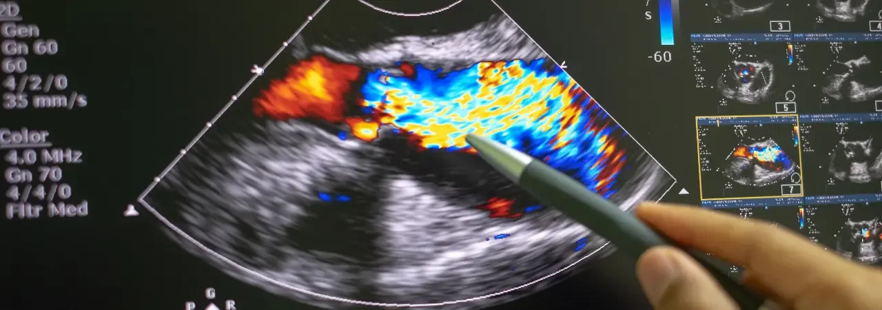
Aort anevrizması ve diseksiyonunda kasıktan girerek damar içinden ilerlediğimiz bu yaklaşımda, stent-greftle yıpranmış bölgeyi içeriden destekleriz. Böylece kanın zayıf duvarla temasını keser, yırtılma riskini hızla azaltırız. Klinik akışta önce tanısal görüntüleme ile plan yapar, ardından uygun cihazı yerleştiririz. Bu çerçevede endovasküler aort tedavisi; hızlı iyileşme, daha az ağrı ve daha kısa hastane yatışı gibi somut faydalar sunar. Uygulamada rehber tel, kılıf ve görüntüleme eşliğinde ilerler, sızdırmazlığı intraoperatif testlerle doğrularız. Ayrıca, uygun endikasyonda evar tevar işlemi güvenli ve etkin bir seçenek haline gelir. Gerektiğinde arkus segmentinde tevar stent greft kullanırız; yaklaşımımız temelde minimal invaziv aort cerrahisi ilkelerine dayanır.
Minimal invaziv aort cerrahisi yaklaşımı
- Önce çok kesitli BT anjiyografi ile anatomiyi netleştiririz.
- Ardından ölçümlerin doğruluğunu teyit eder, uygun iniş/kalkış zonlarını belirleriz.
- Daha sonra erişim yolunu planlar, gerekirse kapalı teknikle kasıktan gireriz.
- Son olarak stent-grefti konumlayıp sızdırmazlık testlerini yapar, hemodinamiği izleriz.
İpucu: Adım adım ilerlemek, kontrast ve radyasyon dozunu optimize etmek kadar önemlidir.
Açık cerrahiye göre avantajlar ve sınırlılıklar
|
|
|
|
|
|
|
|
|
|
|
|
|
|
|
|
|
|
|
|
|
|
|
|
Kısa yol: Ön değerlendirme ve doğru boyutlandırma, komplikasyon oranını belirgin düşürür.
EVAR TEVAR için hasta seçimi ve ön değerlendirme
Aort anevrizması ve diseksiyonu için uygun adayı seçerken, amacımız güvenliği en üstte tutarak başarı oranını artırmaktır. Bu nedenle, planladığımız endovasküler aort tedavisi öncesinde sistematik bir ön değerlendirme uygular, riskleri titizlikle sınıflandırırız. Özellikle Ankara’daki merkezimizde, çok disiplinli kurul ile karar verir; uygun olgularda minimal invaziv aort cerrahisi yaklaşımını tercih ederiz. Böylece, olası komplikasyonları en aza indirip, evar tevar işlemi sürecini öngörülebilir ve hızlı hâle getiririz.
Endikasyonlar, kontraendikasyonlar ve risk sınıflaması
Ön değerlendirmeyi yapılandırılmış bir algoritma ile yürütürüz. İlk adımda endikasyonları doğrular, ardından mutlak/bağıl kontraendikasyonları ve perioperatif riskleri değerlendiririz.
|
|
|
|
|
|
|
|
|
|
|
|
|
|
|
|
|
|
|
|
|
|
|
|
|
|
|
|
|
|
|
|
|
|
|
İpucu: Alerji öyküsünde premedikasyon; antikoagülasyon kullanan hastalarda ilaç yönetimini anestezi ile birlikte planlarız.
BT anjiyografi ile ölçüm ve anatominin uygunluk analizi
BT anjiyografi, planlamanın bel kemiğidir. Önce centerline üzerinden ölçüm alır, sonra iniş alanlarını ve sızdırmazlık için gerekli kapaklanma uzunluklarını belirleriz. Boyun/landing zone uzunluğu, damar çapları, kavis (tortuozite) ve kalsifikasyonu değerlendirir; buna göre tevar stent greft boyutlandırmasını genellikle %10–20 oversize prensibiyle yaparız. Ayrıca:
- İliak/femoral erişimi çap, kalsifikasyon ve şekil olarak doğrularız.
- Visseral dallar, arkus varyantları ve dallanma mesafelerini işaretleriz.
- Gerektiğinde fenestrasyon/branched seçeneklerini ve hibrit yaklaşımları tartışırız.
Son olarak, anestezi stratejisini (lokal + sedasyon ya da genel) ve kontrast/ışın dozunu optimize eder, adım adım planı yazılı hale getiririz; böylece prosedürde sapma olmadan ilerleriz.
evar tevar işlemi adım adım: hazırlık ve anestezi
“Hazırlıkta hedefimiz kanamayı azaltmak, böbreği korumak ve görüntülemeyi kusursuz kılmaktır.”
İşe, kasıktan girişle uygulanan evar tevar işlemi için net, ölçüme dayalı bir planla başlıyoruz. Böylece hem endovasküler aort tedavisi akışını hızlandırıyor hem de komplikasyon riskini azaltıyoruz. Bu yaklaşım, gerçek anlamda minimal invaziv aort cerrahisi felsefesini destekler.
Preoperatif hazırlık, antikoagülasyon ve enfeksiyon profilaksisi
- BT anjio ile aort ve iliak damarların çap/uzunluk/angülasyon ölçümlerini doğrular, olası kapı damar kılavuzlarını planlarız; gerekirse tevar stent greft uyumu için yeniden ölçüm yaparız.
- İlaç yönetimi: Varfarin/DOAC kullanımını kanama ve tromboz riskine göre kesintili veya köprüleme stratejisiyle düzenler, intraoperatif heparinizasyonu ACT hedefleriyle titreriz.
- Böbrek koruma: İzotonik hidrasyon, kontrast miktarını sınırlama, gerekirse NAC ve izo-osmolar kontrast seçimi uygularız.
- Kan yönetimi: Tip-crossmatch, hücre saklama planı ve gerekirse antifibrinolitik strateji belirleriz.
- Enfeksiyon profilaksisi: Zamanında tek doz geniş spektrum profilaksi, klorheksidin banyo ve tıraşsız cilt hazırlığı uygularız.
- Ekip ve ekipman kontrol listesi: Kılavuz teller, kılıflar, kapatma cihazları, balonlar ve yedek giriş planı hazırdır.
- Hastayla onam ve süreç eğitimi: Beklentileri netleştirir, erken mobilizasyonu vurgularız.
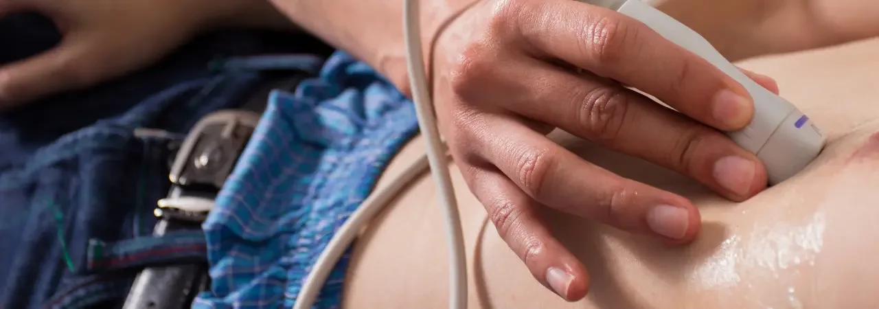
- İpucu: Giriş damar çapı-ultrasonda doğrulama, groin komplikasyonlarını belirgin azaltır.
Anestezi seçenekleri (lokal, rejyonal, genel) ve pozisyonlama
- Pozisyon: Supin, ısı yönetimi, bilateral kasık hazırlığı, floroskopi hattı ve radyasyon koruması optimize edilir.
- Erişim: Ultrason eşliğinde ortak femoral arter ponksiyonu; kapatma cihazı ön yerleştirme (preclose) düşünülebilir.
|
|
|
|
|
|
|
|
|
|
|
|
|
|
|
|
|
|
|
|
Not: Anestezi seçimini hemodinamik durum, akciğer fonksiyonu ve prosedür karmaşıklığına göre kişiselleştiririz; hedefimiz güvenli, ağrısız ve hızlı toparlanmadır.
TEVAR stent greft ve cihaz seçimi: doğru boyutlandırma ve planlama
Doğru cihaz seçimi, görüntülemeyi hassas ölçümlerle birleştirdiğimiz bir planlama disiplinidir. Kontrastlı BT anjiyografide centerline ölçümleriyle çap, uzunluk ve tortuoziteyi belirler; erişim damarlarını önceden değerlendiririz. Böylece evar tevar işlemi sırasında akışı kesmeden güvenli bir yerleşim hedefleriz. Ayrıca tedavi algoritmamızı minimal invaziv aort cerrahisi ilkeleriyle uyumlu tutarız.
Cihaz türleri, kaplama materyalleri ve teslim sistemleri
Seçimi, patoloji segmentine, damar anatomisine ve hedef dallara göre yaparız. Özetle:
|
|
|
|
|
|
|
|
|
|
|
|
|
|
|
|
|
|
|
|
|
|
|
|
Uygun profilde teslim sistemi, kavisli aortta kontrollü ilerleme sağlar; seçtiğimiz tevar stent greft ile sheath-çap uyumunu önceden teyit ederiz. Bu yaklaşım, planlı bir endovasküler aort tedavisi akışı kurmamıza yardım eder.
Landing zone belirleme, oversizing ve seal stratejileri
- Proksimal ve distal landing zone için en az 20–30 mm sağlıklı segment hedefleriz.
- Oversizing’i genellikle %10–20 aralığında tutar, diseksiyonda daha düşük, anevrizmada dengeli seçeriz.
- Seal stratejisinde kötü damar kalitesinde uzatılmış kapama, gerektiğinde LSA yeniden damarlanması ve fenestrasyon/branch seçeneklerini planlarız.
- Karmaşık yay çizgilerinde iğne deliği hizalamasını kolaylaştırmak için işaretli kılavuzlarla çalışır, floroskopi projeksiyonunu önceden belirleriz.
Endovasküler aort tedavisi sırasında prosedür akışı
Bu bölümde, kasıktan ilerlediğimiz kılavuz tel ve kateterlerle süreci adım adım yöneterek evar tevar işlemini güvenle tamamlamayı hedefliyoruz. Amacımız, radyolojik doğrulamalarla ilerleyen, kanama ve emboli riskini azaltan, kanıta dayalı bir akış kurmaktır. Böylece minimal invaziv aort cerrahisi yaklaşımını standardize ederiz.
Giriş yolu seçimi (femoral/iliyak) ve damar hazırlığı
Önce, CTA ile ölçümleri teyit eder; femoral ya da iliyak girişi planlarız. Ultrason eşliğinde ponksiyon yapar, kılavuz tel ve kılıfı yerleştiririz. Heparinizasyon dozunu ağırlığa göre uygular, basınçlı yıkama ve hava boşaltma prosedürleriyle seti hazırlarız. Ardından, referans işaretleri için pigtail kateterle aortografiyi gerçekleştirir, iniş ve çıkış zonlarını işaretleriz. İhtiyaç halinde pre-dilatasyon yaparız. Bu adımlar, akışın sorunsuz ilerlemesi için kritiktir.
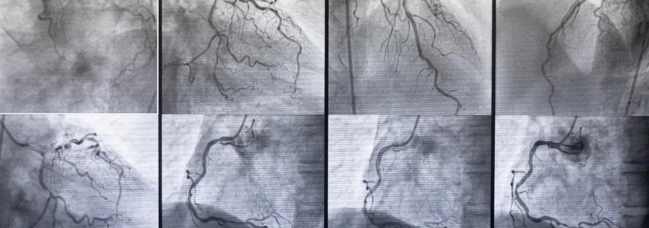
Stent greft implantasyonu, görüntüleme ile doğrulama ve final kontrol
Sonrasında, planlanan konfigürasyonda tevar stent greft yerleştiririz. Floroskopi ve road-map ile doğru seviyeyi hizalar, solunum-apne ve tansiyon kontrolü altında grefti açarız. Gerekirse post-dilatasyon balonlama yaparız. Nihai aortografiyle sızdırmazlığı (endo-lik) kontrol eder, visseral ve iliak akımın korunduğunu doğrularız. Son olarak kapama cihazlarıyla kanülasyon yerini güvenceye alırız ve hemodinamik-stent pozisyon takibini başlatırız. Bu sistematik yaklaşım, endovasküler aort tedavisi başarısını artırır.
|
|
|
|
|
|
|
|
|
|
|
|
|
|
|
|
|
|
|
|
|
|
|
|
Not: İlaçlar, cihaz çapları ve uzunlukları hasta anatomisine göre özelleştirilmelidir; planı, görüntüleme bulgularına göre gerçek zamanlı optimize ederiz.
Komplikasyonların önlenmesi ve yönetimi
Ankara’da uyguladığımız risk odaklı yaklaşım ile komplikasyonları en baştan azaltmayı hedefliyoruz. Bu amaçla görüntüleme ile ayrıntılı planlama yapıyor, damar erişimini ölçümlendiriyor ve uygun tevar stent greft seçimini titizlikle sağlıyoruz. Böylece hem sızıntı hem de istenmeyen migrasyon riskini düşürüyoruz. Ayrıca protokollerimiz, hemodinami ve pıhtılaşma takibini standartlaştırır; gerektiğinde hızlı müdahale için hibrit salon hazırlığı yaparız. Unutmayalım, evar tevar işlemi özünde minimal invaziv aort cerrahisi yaklaşımının güvenli prensipleriyle ilerlediğinde en iyi sonucu verir ve bütün süreç, kanıta dayalı endovasküler aort tedavisi stratejileriyle yönetilir.
“Her adımı ölç, her riski önceden düşün, komplikasyon ortaya çıkarsa gecikmeden düzelt.”
Endoleak tipleri (I–V) ve tedavi yaklaşımları
|
|
|
|
|
|
|
|
|
|
|
|
|
|
|
|
|
|
|
|
|
|
|
|
|
|
|
|
|
|
Böbrek fonksiyonu, nörolojik ve distal iskemi risklerinin azaltılması
- Adım 1: Kontrastı sınırla; izotonik hidrasyon ve nefropati profilaksisi uygularız.
- Adım 2: Spinal perfüzyonu korumak için kan basıncını hedef aralıkta tutar, uzun segment kapatmadan kaçınırız.
- Adım 3: Distal emboliyi azaltmak için filtre, nazik tel-kateter tekniği ve dikkatli pıhtı yönetimi kullanırız.
- Adım 4: Erken dönem Doppler ve kreatinin takibi ile sorunları hızla saptar, gerektiğinde endovasküler rötuş veya yoğun bakım destek protokollerini devreye alırız.
Ankara’da evar tevar sonrası bakım ve takip
Taburculuk kriterleri, ilaç yönetimi ve yaşam tarzı önerileri
Taburculuk öncesi net bir plan yaparız. Kriterlerimiz: stabil vital bulgular, kasık giriş yerinde kanama olmaması, ağrı kontrolü, bağımsız mobilizasyon ve idrar çıkışının normal olması. Ayrıca, evar tevar işlemi sonrası reçete ve eğitimleri adım adım açıklarız.
- İlaçlar: antiplatelet, tansiyon düzenleyici ve gerekirse statin; doz ve etkileşimleri yazılı veririz.
- Yaşam tarzı: sigarayı bırakır, tuz kısıtlı diyet ve düzenli yürüyüş öneririz.
- Basınç hedefi: evde tansiyon takibini öğretir, hedefleri netleriz.
- Giriş yeri bakımı: her gün kontrol, kızarıklık/akıntı varsa bizi arama.
Ankara’da randevu ve hızlı değerlendirme için aynı hafta içi kontrol penceresi sunarız. İhtiyaç halinde, minimal invaziv aort cerrahisi ekibiyle multidisipliner yönetim planları oluştururuz.
İzlem programı: kontrol görüntülemeleri ve alarm bulguları
Ankara’daki merkezimizde takip planını baştan planlarız. İlk ay, 6. ay ve 12. ayda BT anjiyo/dupleks ultrason; ardından yıllık izlem uygularız. Gerektiğinde endovasküler aort tedavisi sonrası ince ayarları hızla yapar, tevar stent greft konumunu ve sızdırmazlığı dikkatle değerlendiririz.
Aşağıdaki tablo, uyguladığımız standart izlem akışını özetler:
|
|
|
|
|
|
|
|
|
|
|
|
|
|
|
|
|
|
|
|
|
|
|
|
|
|
|
|
|
|
İpuçları:
- Randevu öncesi bol su için; kontrast nefropatisini önlemek için böbrek fonksiyon testlerini birlikte planlarız.
- Antiplatelet kesintisi gerekirse, cerrahi/dental işlemlerden önce bizi arayın; en güvenli aralığı birlikte belirleyelim.
- Evde tansiyon ve nabız ölçümlerinizi haftalık olarak kaydedin; sapmaları bize iletin.